Short answer
Axonal outputs are coupled to dendrites or other effector tissues, such as muscular fibers or glandular tissue. There are no redundant axonal outputs, or redundant dendritic inputs. There is a tight coupling between the two both during developmental formation of synapses and during the maintenance of the two.
Background
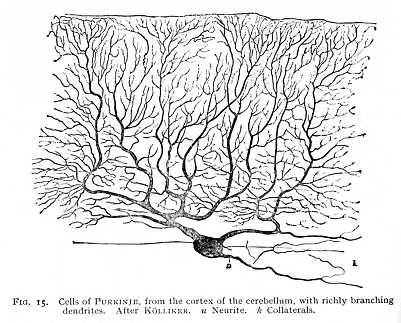
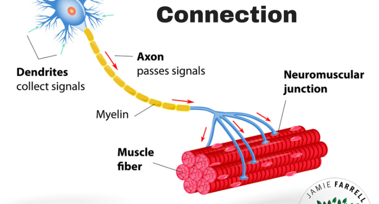
Neurons can have extensive dendritic trees (e.g., Purkinje cells in the cerebellar cortex, Fig. 1) and also elaborate arborated axon terminals (*e.g., in the neuromuscular junction, Fig. 2).
In the cases shown below, the functions can be defined as simple logical operators, namely integration (Purkinje cell) and amplification (neuromuscular junction). The Purkinje cells receive and integrate input from the brainstem. The motoneurons innervating muscle use multiple axon terminals to target a larger area of the muscle so that stronger, synchronous muscle contractions can be achieved.
The development and maintenance of dendrite-axon connections are tightly coupled (e.g., Shen & Lowan, 2010). Generally, when either input to the dendrite is diminished (axonal regression), or output of the axon is cancelled (dendritic regression), the plasticity of the neural tissue will lead to degeneration or re-directing of the axon, or regression or re-innervation of the dendrite, respectively (e.g. Marc et al., 2003).
As regarding to your math; be aware axons can branch, axon arborations can target a single dendritic tree, axons can terminate on other tissues, such as glands and muscles (no dendrites there!). In other words, your mathematical example is an oversimplification. Further; cells with elaborate dendritic trees as shown in Fig. 1 are common in cortical layers, but are not the norm. The ratios of the cell types have to be taken into account as well.
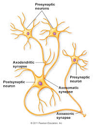
Lastly, there can be the textbook connections between neurons, namely axo-dendritic connections, but also axo-axonal (Schmitz et al, 2001), dendrodendritic (Shepherd, 2009), and even axosomatic synapses between neurons (Fig. 3).

Fig. 1. Dendritic tree (Wundt, 1904)

Fig. 2. Axonal terminals in a neuromuscular junction. source: Farrel (2017)

Fig. 3. Different kinds of synapses. source: Modesto Junior College
References
- Marc et al., Prog Retin Eye Res (2003); 22(5):607-55
- Shen & Kowan, Cold Spring Harb Perspect Biol (2010); 2(4): a001842
- Shepherd, Ann N Y Acad Sci (2009); 1170; 1-11
- Schmitz et al., Neuron (2001); 31(5): 831-40
- Wundt, Principles of Physiological Psychology (1904)


