Do visual cortical signals always go back and forth between the thalamus, or other subcortical structures, or can they travel directly from one region in the cortex to another?
-
$\begingroup$ cortical integration of wernicke's and broca's area: auditory input are processed in wernicke's then it is transposed to the broca's area for motor processes involved in speech. the thalamus is bypassed in this example. $\endgroup$– Christopher S ParkCommented Sep 21, 2016 at 7:46
-
$\begingroup$ I've restricted the question to the visual system just for the sake of narrowing down the question. According to our dialogue an example is sufficient for you anyway and the question was too broad according to the conventions of this site. $\endgroup$– AliceD ♦Commented Feb 18, 2017 at 20:15
1 Answer
Short answer
Intracortical projections can be routed directly to other cortical areas (cortico-cortical projections), or via the thalamus (cortico-thalamo-cortical projections).
Background
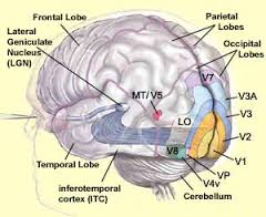
Intracortical projections do not have to cross the thalamus or other subcortical structures. Take for example the visual system - it features many cortical projections and these connections do not always enter the thalamus or other subcortical structures (Fig. 1). These connections are called cortico-cortical projections (e.g., Guandalani, 1998). Connections going via the thalamus, however, also exist in the visual cortex (e.g., Kato, 1990) and are referred generally to as cortico-thalamo-cortical projections.

Fig. 1. Visual system. source: McGill
The projections from the LGN to primary cortex (V1 in Fig. 1) and via V2, V3, V4 up to V5 all may occur without visual information entering the thalamus or other subcortical structures.
Also note that on the cellular scale direct cortico-cortical connections in the cortex are abundant (Fig. 2).

Fig. 2. Local cellular connections in the visual cortex. source: Kang et al. (2014)
Reference
- Guandalani, Brain Res Bull; 47(4): 377–85
- Kang et al., Front Syst Neurosci (2014); 8: 172
- Kato, Brain Res; 509(1): 150–2
-
$\begingroup$ Do you know why on Fig2 B the big green arrow is going up the apical dendrite? $\endgroup$– ceillacCommented Feb 20, 2017 at 5:16