Commonmark migration
Source Link

Based on the most recent (all post-2009) research, it seems to be the same general pathway, just with different gradients in the same areas of the brain. From Taylor et al. (2016):

Recent studies suggest that dopamine neurons in the VTA [ventral tegmental area] and SN [substantia nigra] form a heterogeneous population tuned to either (or both) aversive or rewarding stimuli.


The heterogeneity of dopamine neurons in response to aversive and rewarding stimuli suggests that they serve unique functional roles. Cells activated by reward and inhibited by punishment are well suited to code motivational valence, whereas neurons activated by both rewarding and punishing stimuli are likely to code motivational salience [stimulus awareness]. Neurons coding motivational valence [whether the stimulus is positive or negative in value] would provide a signal for reward seeking, evaluation, and value learning, in line with current theories on the role of dopamine in reward processing. In contrast, neurons coding motivational salience would provide a signal for detection and prediction of highly important events independent of valence, pursuant to dopamine's role in salience processing. These distinct aspects of dopamine neurotransmission might be neuroanatomically separate: dopaminergic neurons coding motivational valence have been found more commonly in the ventromedial SN and lateral VTA with projections to nucleus accumbens [NAc] shell, whereas neurons coding motivational salience are more often reported in the dorsolateral SN with projections to the nucleus accumbens core.


enter image description here

The primary evidence for this updated view seems mostly based (in order of the number of citations) on 3 papers:


The part of your question that probably confused Alice and also confuses me a little is that you also ask (in your final paragraph) about somatosensory mapping. The latter doesn't generally involve tracing any pathways beyond the sensory areas of the brain, e.g. for macaques

enter image description here

Also such maps are not totally fixed as there is some use-dependent plasticity.
Based on the most recent (all post-2009) research, it seems to be the same general pathway, just with different gradients in the same areas of the brain. From Taylor et al. (2016):

Recent studies suggest that dopamine neurons in the VTA [ventral tegmental area] and SN [substantia nigra] form a heterogeneous population tuned to either (or both) aversive or rewarding stimuli.

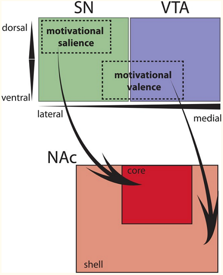
The heterogeneity of dopamine neurons in response to aversive and rewarding stimuli suggests that they serve unique functional roles. Cells activated by reward and inhibited by punishment are well suited to code motivational valence, whereas neurons activated by both rewarding and punishing stimuli are likely to code motivational salience [stimulus awareness]. Neurons coding motivational valence [whether the stimulus is positive or negative in value] would provide a signal for reward seeking, evaluation, and value learning, in line with current theories on the role of dopamine in reward processing. In contrast, neurons coding motivational salience would provide a signal for detection and prediction of highly important events independent of valence, pursuant to dopamine's role in salience processing. These distinct aspects of dopamine neurotransmission might be neuroanatomically separate: dopaminergic neurons coding motivational valence have been found more commonly in the ventromedial SN and lateral VTA with projections to nucleus accumbens [NAc] shell, whereas neurons coding motivational salience are more often reported in the dorsolateral SN with projections to the nucleus accumbens core.

enter image description here

The primary evidence for this updated view seems mostly based (in order of the number of citations) on 3 papers:


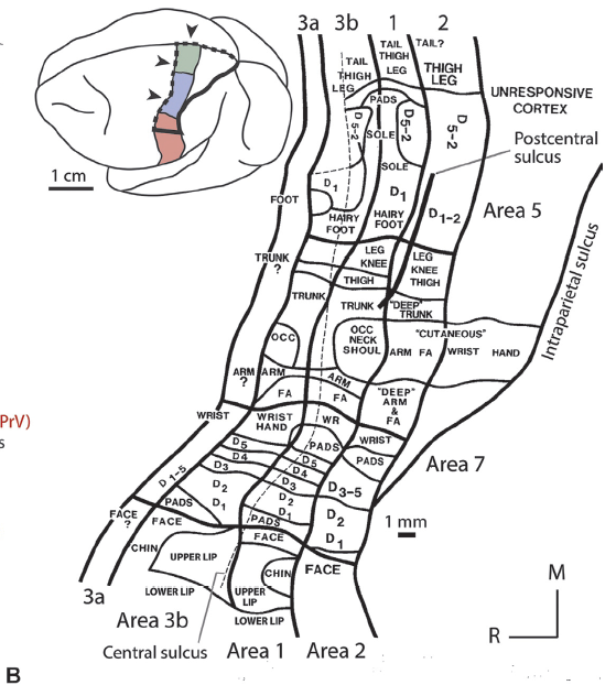
The part of your question that probably confused Alice and also confuses me a little is that you also ask (in your final paragraph) about somatosensory mapping. The latter doesn't generally involve tracing any pathways beyond the sensory areas of the brain, e.g. for macaques

enter image description here

Also such maps are not totally fixed as there is some use-dependent plasticity.

since a physical length is given in the figure
Source Link

Based on the most recent (all post-2009) research, it seems to be the same general pathway, just with different gradients in the same areas of the brain. From Taylor et al. (2016):

Recent studies suggest that dopamine neurons in the VTA [ventral tegmental area] and SN [substantia nigra] form a heterogeneous population tuned to either (or both) aversive or rewarding stimuli.

The heterogeneity of dopamine neurons in response to aversive and rewarding stimuli suggests that they serve unique functional roles. Cells activated by reward and inhibited by punishment are well suited to code motivational valence, whereas neurons activated by both rewarding and punishing stimuli are likely to code motivational salience [stimulus awareness]. Neurons coding motivational valence [whether the stimulus is positive or negative in value] would provide a signal for reward seeking, evaluation, and value learning, in line with current theories on the role of dopamine in reward processing. In contrast, neurons coding motivational salience would provide a signal for detection and prediction of highly important events independent of valence, pursuant to dopamine's role in salience processing. These distinct aspects of dopamine neurotransmission might be neuroanatomically separate: dopaminergic neurons coding motivational valence have been found more commonly in the ventromedial SN and lateral VTA with projections to nucleus accumbens [NAc] shell, whereas neurons coding motivational salience are more often reported in the dorsolateral SN with projections to the nucleus accumbens core.

enter image description here

The primary evidence for this updated view seems mostly based (in order of the number of citations) on 3 papers:


The part of your question that probably confused Alice and also confuses me a little is that you also ask (in your final paragraph) about somatosensory mapping. The latter doesn't generally involve tracing any pathways beyond the sensory areas of the brain, e.g. for mammalsmacaques

enter image description here

Also such maps are not totally fixed as there is some use-dependent plasticity.

Based on the most recent (all post-2009) research, it seems to be the same general pathway, just with different gradients in the same areas of the brain. From Taylor et al. (2016):

Recent studies suggest that dopamine neurons in the VTA [ventral tegmental area] and SN [substantia nigra] form a heterogeneous population tuned to either (or both) aversive or rewarding stimuli.

The heterogeneity of dopamine neurons in response to aversive and rewarding stimuli suggests that they serve unique functional roles. Cells activated by reward and inhibited by punishment are well suited to code motivational valence, whereas neurons activated by both rewarding and punishing stimuli are likely to code motivational salience [stimulus awareness]. Neurons coding motivational valence [whether the stimulus is positive or negative in value] would provide a signal for reward seeking, evaluation, and value learning, in line with current theories on the role of dopamine in reward processing. In contrast, neurons coding motivational salience would provide a signal for detection and prediction of highly important events independent of valence, pursuant to dopamine's role in salience processing. These distinct aspects of dopamine neurotransmission might be neuroanatomically separate: dopaminergic neurons coding motivational valence have been found more commonly in the ventromedial SN and lateral VTA with projections to nucleus accumbens [NAc] shell, whereas neurons coding motivational salience are more often reported in the dorsolateral SN with projections to the nucleus accumbens core.

enter image description here

The primary evidence for this updated view seems mostly based (in order of the number of citations) on 3 papers:


The part of your question that probably confused Alice and also confuses me a little is that you also ask (in your final paragraph) about somatosensory mapping. The latter doesn't generally involve tracing any pathways beyond the sensory areas of the brain, e.g. for mammals

enter image description here

Based on the most recent (all post-2009) research, it seems to be the same general pathway, just with different gradients in the same areas of the brain. From Taylor et al. (2016):

Recent studies suggest that dopamine neurons in the VTA [ventral tegmental area] and SN [substantia nigra] form a heterogeneous population tuned to either (or both) aversive or rewarding stimuli.

The heterogeneity of dopamine neurons in response to aversive and rewarding stimuli suggests that they serve unique functional roles. Cells activated by reward and inhibited by punishment are well suited to code motivational valence, whereas neurons activated by both rewarding and punishing stimuli are likely to code motivational salience [stimulus awareness]. Neurons coding motivational valence [whether the stimulus is positive or negative in value] would provide a signal for reward seeking, evaluation, and value learning, in line with current theories on the role of dopamine in reward processing. In contrast, neurons coding motivational salience would provide a signal for detection and prediction of highly important events independent of valence, pursuant to dopamine's role in salience processing. These distinct aspects of dopamine neurotransmission might be neuroanatomically separate: dopaminergic neurons coding motivational valence have been found more commonly in the ventromedial SN and lateral VTA with projections to nucleus accumbens [NAc] shell, whereas neurons coding motivational salience are more often reported in the dorsolateral SN with projections to the nucleus accumbens core.

enter image description here

The primary evidence for this updated view seems mostly based (in order of the number of citations) on 3 papers:


The part of your question that probably confused Alice and also confuses me a little is that you also ask (in your final paragraph) about somatosensory mapping. The latter doesn't generally involve tracing any pathways beyond the sensory areas of the brain, e.g. for macaques

enter image description here

Also such maps are not totally fixed as there is some use-dependent plasticity.

added 405 characters in body
Source Link

Based on the most recent (all post-2009) research, it seems to be the same general pathway, just with different gradients in the same areas of the brain. From Taylor et al. (2016):

Recent studies suggest that dopamine neurons in the VTA [ventral tegmental area] and SN [substantia nigra] form a heterogeneous population tuned to either (or both) aversive or rewarding stimuli.

The heterogeneity of dopamine neurons in response to aversive and rewarding stimuli suggests that they serve unique functional roles. Cells activated by reward and inhibited by punishment are well suited to code motivational valence, whereas neurons activated by both rewarding and punishing stimuli are likely to code motivational salience [stimulus awareness]. Neurons coding motivational valence [whether the stimulus is positive or negative in value] would provide a signal for reward seeking, evaluation, and value learning, in line with current theories on the role of dopamine in reward processing. In contrast, neurons coding motivational salience would provide a signal for detection and prediction of highly important events independent of valence, pursuant to dopamine's role in salience processing. These distinct aspects of dopamine neurotransmission might be neuroanatomically separate: dopaminergic neurons coding motivational valence have been found more commonly in the ventromedial SN and lateral VTA with projections to nucleus accumbens [NAc] shell, whereas neurons coding motivational salience are more often reported in the dorsolateral SN with projections to the nucleus accumbens core.

enter image description here

The primary evidence for this updated view seems mostly based (in order of the number of citations) on 3 papers:


The part of your question that probably confused Alice and also confuses me a little is that you also ask (in your final paragraph) about somatosensory mapping. The latter doesn't generally involve tracing any pathways beyond the sensory areas of the brain, e.g. for mammals

enter image description here

Based on the most recent (all post-2009) research, it seems to be the same general pathway, just with different gradients in the same areas of the brain. From Taylor et al. (2016):

Recent studies suggest that dopamine neurons in the VTA [ventral tegmental area] and SN [substantia nigra] form a heterogeneous population tuned to either (or both) aversive or rewarding stimuli.

The heterogeneity of dopamine neurons in response to aversive and rewarding stimuli suggests that they serve unique functional roles. Cells activated by reward and inhibited by punishment are well suited to code motivational valence, whereas neurons activated by both rewarding and punishing stimuli are likely to code motivational salience [stimulus awareness]. Neurons coding motivational valence [whether the stimulus is positive or negative in value] would provide a signal for reward seeking, evaluation, and value learning, in line with current theories on the role of dopamine in reward processing. In contrast, neurons coding motivational salience would provide a signal for detection and prediction of highly important events independent of valence, pursuant to dopamine's role in salience processing. These distinct aspects of dopamine neurotransmission might be neuroanatomically separate: dopaminergic neurons coding motivational valence have been found more commonly in the ventromedial SN and lateral VTA with projections to nucleus accumbens [NAc] shell, whereas neurons coding motivational salience are more often reported in the dorsolateral SN with projections to the nucleus accumbens core.

enter image description here

The primary evidence for this updated view seems mostly based (in order of the number of citations) on 3 papers:


The part of your question that probably confused Alice and also confuses me a little is that you also ask (in your final paragraph) about somatosensory mapping. The latter doesn't generally involve tracing any pathways beyond the sensory areas of the brain, e.g. for mammals

enter image description here

Source Link
Loading